Co-reporter:Chenjuan Zhou, Xue Sun, Jing Yan, Binhe Chen, Peiran Li, Huigang Wang, Jiyang Liu, Xiaoping Dong, Fengna Xi
Powder Technology 2017 Volume 308() pp:114-122
Publication Date(Web):15 February 2017
DOI:10.1016/j.powtec.2016.11.056
•Dyes can be decomposed by the thermo-driven catalysis of g-C3N4.•A synergetic effect of g-C3N4 and H2O2 is demonstrated.•This approach shows the superior applicability and reusability.•The doping level promotes the separation of thermo-generated e−–h+.Herein, graphitic carbon nitride (g-C3N4) as a novel thermo-driven catalyst was used for catalytic decomposition of organic dyes with the assistance of H2O2. In comparison with the experiment with individual catalyst or H2O2, a prominent enhancement of performance was achieved by the synergetic effect of g-C3N4 and H2O2. Experimental parameters like the H2O2 dosage and reaction temperature were optimized, and the catalyst exhibited excellent application universality for various concentration and different dyes. By physicochemical characterizations, the modification or doping with oxygen-containing groups on carbon nitride framework was demonstrated. We proposed a possible mechanism that the doping level trapped thermo-excited electrons to promote the separation of electrons and holes. On the other hand H2O2 was reduced by the excited electron to produce hydroxyl radical. The quenching experiment suggested the holes and hydroxyl radical were the main active species, thus verifying the synergetic effect of g-C3N4 and H2O2. Additionally, the superior stability and reusability were also proved by the successive five cycles, where the activity was even gradually improved with the increase of cycle number.
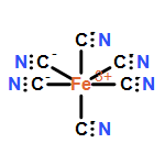
Co-reporter:Xue Sun, Yuting Qian, Yajie Jiao, Jiyang Liu, Fengna Xi, Xiaoping Dong
Talanta 2017 Volume 165() pp:429-435
Publication Date(Web):1 April 2017
DOI:10.1016/j.talanta.2016.12.085
•Ionic liquid-capped GQDs (IL-GQDs) were easily prepared.•IL-GQDs was used as label-free fluorescent probe for direct detection of Fe(CN)63−.•Sensitive detection of Fe(CN)63− with low detection limit was achieved.Despite complex molecular and atomic doping, efficient post-functionalization strategies for graphene quantum dots (GQDs) are of key importance to control the physicochemical properties and broaden the practical applications. With ionic liquid as specific modification agents, herein, the preparation of ionic liquid-capped GQDs (IL-GQDs) and its application as label-free fluorescent probe for direct detection of anion were reported. Hydroxyl-functionalized GQDs that could be easily gram-scale synthesized and possessed single-crystalline were chosen as the model GQDs. Also, the most commonly used ionic liquids, water-soluble 1-butyl-3-methyl imidazolium tetrafluoroborate (BMIMBF4) was chosen as the model IL. Under the ultrasonic treatment, BMIMBF4 easily composited with GQDs to form IL-GQDs. The synthesized IL-GQDs were characterized by atomic force microscopy (AFM), transmission electron microscopy (TEM), X-ray photoelectron spectroscopy (XPS) and fluorescence (FL) spectrum. After successful combination with IL, the excitation-independent photoluminescence behavior of GQDs presented almost no change, whereas, the anion responsiveness of IL-GQDs drastically improved, which afforded the IL-GQDs a sensitive response to Fe(CN)63−. Based on the strong fluorescence quench, a facile and sensitive detection of Fe(CN)63− was achieved. A wide linear range of 1.0×10⁻⁷ to 2.5×10⁻³ mol l−1 with a low detection limit of 40 nmol l−1 was obtained. As the composition and properties of IL and GQDs could be easily tuned by varying the structure, ionic liquids-capped GQDs might present promising potential for their applications in sensing and catalysis.
Co-reporter:Shiyue Bian, Chao Shen, Yuting Qian, Jiyang Liu, Fengna Xi, Xiaoping Dong
Sensors and Actuators B: Chemical 2017 Volume 242() pp:231-237
Publication Date(Web):April 2017
DOI:10.1016/j.snb.2016.11.044
•One-step synthesis of S-GQDs was developed.•Successful doping of S atom in GQDs was proven.•S-GQDs exhibited monolayer-graphene thickness and high crystallinity.•Facile and direct fluorescence sensor for sensitive detection of Ag+ was achieved.Sulfur-doped graphene quantum dots (S-GQDs) with bright blue emission have been prepared by a facile one-pot hydrothermal treatment. A specific compound, 1,3,6-trinitropyrene, which has a mother nucleus structure similar with graphene, was chosen as the carbon source and 3-mercaptopropionic acid (MPA) was employed for S-doping and carboxyl groups modification. The synthesized S-GQDs were characterized by atomic force microscopy (AFM), transmission electron microscope (TEM), X-ray photoelectron spectroscopy (XPS) and fluoscence (FL) spectrum. Results indicated that S-GQDs possessed single layer graphene structure with mean size of about 2.5 nm and presented an excitation-independent photoluminescence behavior with maximum excitation/emission wavelength at 360/450 nm, respectively. The sulfur-doping of GODs drastically improved their electronic and chemical properties, which afforded the S-GQDs a sensitive response to Ag+ ions. Furthermore, the S-GQDs were successfully explored as a sensing probe for Ag+ detection with high sensitivity and selectivity. A wide linear range of 0.1-130.0 μM with a low detection limit of 30 nM was obtained. The facile preparation method and the high performace of the as-prepared S-GQDs present promising potential for their applications in sensing, biological imaging and catalysis.
Co-reporter:Jing Yan, Xiaoxue Han, Xiaozhong Zheng, Jiajia Qian, Jiyang Liu, Xiaoping Dong, Fengna Xi
Materials Research Bulletin 2017 Volume 94(Volume 94) pp:
Publication Date(Web):1 October 2017
DOI:10.1016/j.materresbull.2017.06.022
•The synthetic approach offers the merits of low-cost and high efficiency.•The PCNF materials are porous and ultrathin.•The PCNF materials exhibit enhanced photocatalytic performance.•This work provides a method to synthesize other porous two-dimensional materials.A facile one-step approach was employed to synthesize porous g-C3N4 flake (PCNF) via thermally polymerizing melamine with the presence of MgCO3 template. CO2 gas released from the decomposition of MgCO3 prohibited the further polymerization of CN framework and separated the CN layers to produce a flake-like morphology, and meanwhile the removal of MgO nanoparticles resulted in a porous structure. The resultant PCNF exhibited enhanced visible-light photocatalytic activity for degrading organic dyes, and its rate constant is ∼4 times higher than that of bulky CN. What is more, this material presented an excellent stability and superior universality for various pollutants.Download high-res image (206KB)Download full-size image
Co-reporter:Mingfeng Chen, Huaqing Xuan, Xiaozhong Zheng, Jiyang Liu, Xiaoping Dong, Fengna Xi
Electrochimica Acta 2017 Volume 238(Volume 238) pp:
Publication Date(Web):1 June 2017
DOI:10.1016/j.electacta.2017.04.034
Well-controlled mesoporosity is of importance for porous carbons as electrochemical electrode materials. However, the ordered mesoporous carbons prepared from the template approaches face the fact of relative low specific surface area in comparison to activated carbons. Herein, we employed a hard-template route associated with the chemical activation to prepare N-doped mesoporous carbon by co-casting of carbon and nitrogen precursors into the pore channels of mesoporous silica. The obtained activated N-doped mesoporous carbon (ANMC) material preserved the morphology and mesoporous structure of template, and meanwhile a secondary mesoporosity was introduced by the KOH activation. It was demonstrated that the dominant porosity in ANMC sample was from mesopore, and it possessed a high mesopore surface area (2505.6 m2 g−1) and mesopore volume (1.74 cm3 g−1). The N dopant was determined to be pyridinic-N, pyrrolic-N, quaternary-N and pyridine-N-oxide, which did not only contribute the pseudocapaticance but also facilitated the electron transfer in the carbon skeleton. The developed mesoporosity and N doping made this material exhibit superior electrochemical performance that was much higher than those of ordered mesoporous carbon, N-doped ordered mesoporous carbon and activated N-doped carbon samples. Also, the specific capacitance 336.9 F g−1 (0.5 A g−1) and the rate capability were higher than those of other reported mesoporous carbons. In addition, the assembled symmetrical supercapaictor simultaneously showed the high energy density and power density, as well as presented the superb cycling ability (∼98.5% capacitance retaining after 5000 runs).Download high-res image (184KB)Download full-size image
Co-reporter:Chao Shen;Shuyan Ge;Youyou Pang;Jiyang Liu;Xiaoping Dong;Peng Chen
Journal of Materials Chemistry B 2017 vol. 5(Issue 32) pp:6593-6600
Publication Date(Web):2017/08/16
DOI:10.1039/C7TB00506G
Sensitive and selective detection of metal ions is important for environmental monitoring and biological studies because metal ions play crucial roles in many physiological and pathological processes including cellular metabolism, enzymatic activities, as well as DNA and RNA syntheses. In this work, new nitrogen and sulfur co-doped graphene quantum dots (N,S-GQDs) are synthesized and serve as fluorescence probes for parallel and specific detection of Fe3+, Cu2+ and Ag+ ions. The N,S-GQDs are synthesized based on one-step bottom-up molecular fusion in a hydrothermal process with a high yield of 87.8% and possibility of mass production. The as-prepared N,S-GQDs exhibit a single-layer graphene structure, high crystallinity and uniform size (∼2.1 nm). Successful co-doping of N and S atoms in the lattice of GQDs not only enables bright blue fluorescence with high absolute photoluminescence quantum yield (23.2%) but also provides unique selectivity towards some metal ions. With the help of different masking agents, N,S-GQDs are able to differentially detect Fe3+, Cu2+ and Ag+ ions in a mixture with low detection limits (8 nM, 250 nM and 50 nM, respectively). Moreover, detection of Fe3+, Cu2+ and Ag+ in complex biological (serum) and environmental samples (river water) is demonstrated.
Co-reporter:Cong Qiumei, Bian Hongmei, Yu Zhaoxia, Jiyang Liu and Fengna Xi
Analytical Methods 2015 vol. 7(Issue 22) pp:9655-9662
Publication Date(Web):19 Oct 2015
DOI:10.1039/C5AY01871D
Facile electrochemical methods for highly sensitive detection of tumor markers provide great advances in early clinical diagnosis of cancer and public health protection. Herein, a reagentless electrochemical immunosensing platform was developed for sensitive immunoassay of tumor biomarkers based on surface-confined probes and the layer-by-layer assembly technique. Ferrocene grafted cationic polymer polyethyleneimine (PEI–Fc) was modified on chemically reduced graphene oxide (rGO) to form a redox-active and positively charged PEI–Fc–G nanocomposite. Through the layer-by-layer electrostatic assembly technique, the positively charged PEI–Fc–G and negatively charged anionic polyelectrolyte sodium-p-styrene (PSS) were alternately assembled on a negatively charged Au electrode. Based on the biospecific binding of lectin and sugarprotein, the concanavalin A (Con A) lectin monolayer served as the linker to immobilize sugarprotein (horseradish peroxidase, HRP) labeled anti-CEA antibody (HRP-Ab) on the surface of the (PEI–Fc–G/PSS)n/PEI multilayer substrate. With carcinoembryonic antigen (CEA) being the model tumor biomarker, the as-prepared immunosensor presented high selectivity and good stability for sensitive and reagentless detection of CEA with a wide range of 0.1 ng mL−1 to 120 ng mL−1 (R2 = 0.9963) and a detection limit as low as 60 pg mL−1 at a signal/noise ratio of 3. The proposed immunosensor might serve as a versatile platform for reliable cancer diagnostics clinical and biochemical analysis.
Co-reporter:Jiyang Liu, Jiao Wang, Tianshu Wang, Dan Li, Fengna Xi, Jin Wang, Erkang Wang
Biosensors and Bioelectronics 2015 Volume 65() pp:281-286
Publication Date(Web):15 March 2015
DOI:10.1016/j.bios.2014.10.016
•Immunosensor based on monolithic 3D graphene was prepared for the first time.•3D graphene was functionalized by in-situ polymerization of dopamine.•Polydopamine layer imparts 3D graphene with well hydrophilicity and modifiability.•Lectin grafted on polydopamine was used to immobilize sugar-protein labeled Ab.•A wide detection range of 0.1–750.0 ng ml−1 was reached.A high performance three-dimensional (3D) electrochemical immunosensor was developed for sensitive detection of the tumor biomarker, carcinoembryonic antigen (CEA). Monolithic and macroporous graphene foam grown by chemical vapor deposition (CVD) served as the scaffold of the free-standing 3D electrode. Immuno-recognition interface was fabricated via simple and non-covalent immobilization of antibody using lectin-mediated strategy. Briefly, the well-known lectin macromolecule (concanavalin A, Con A) monolayer was functionalized on 3D graphene (3D-G) using in-situ polymerized polydopamine as the linker. Then the widely used horseradish peroxidase (HRP)-labeled antibody (anti-CEA) in immunoassays was efficiently immobilized to demonstrate the recognition interface via the biospecific affinity of lectin with sugarprotein. The 3D immunosensor is able to detect CEA with a wide linear range (0.1–750.0 ng ml−1), low detection limit (~90 pg ml−1 at a signal-to-noise ratio of 3), and short incubation time (30 min). Furthermore, this biosensor was used for the detection of the CEA level in real serum samples.
Co-reporter:Jiyang Liu, Xiaohui Wang, Tianshu Wang, Dan Li, Fengna Xi, Jin Wang, and Erkang Wang
ACS Applied Materials & Interfaces 2014 Volume 6(Issue 22) pp:19997
Publication Date(Web):November 10, 2014
DOI:10.1021/am505547f
Biological modification of monolithic and porous 3D graphene is of great significance for extending its application in fabricating highly sensitive biosensors. The present work reports on the first biofunctionalization of monolithic and freestanding 3D graphene foam for one-step preparation of reagentless enzymatic biosensors by controllable chitosan (CS) electrodeposition technology. Using a homogeneous three-component electrodeposition solution containing a ferrocene (Fc) grafted CS hybrid (Fc-CS), glucose oxidase (GOD), and single-walled carbon nanotubes (SWNTs), a homogeneous biocomposite film of Fc-CS/SWNTs/GOD was immobilized on the surface of 3D graphene foam by one-step electrodeposition. The Fc groups grafted on chitosan can be stably immobilized on the 3D graphene surface and keep their original electrochemical activity. The SWNTs doped into the Fc-CS matrix act as a nanowire to facilitate electron transfer and improve the conductivity of the biocomposite film. Combined with the extraordinary properties of 3D graphene foam including large active surface area, high conductivity, and fast mass transport dynamics, the 3D graphene based enzymatic biosensor achieved a large linear range (5.0 μM to 19.8 mM), a low detection limit (1.2 μM), and rapid response (reaching the 95% steady-state response within 8 s) for reagentless detection of glucose in the phosphate buffer solution.Keywords: 3D graphene; biosensor; chitosan electrodeposition; enzyme; reagentless
Co-reporter:
Analytical Methods (2009-Present) 2015 - vol. 7(Issue 22) pp:NaN9662-9662
Publication Date(Web):2015/10/19
DOI:10.1039/C5AY01871D
Facile electrochemical methods for highly sensitive detection of tumor markers provide great advances in early clinical diagnosis of cancer and public health protection. Herein, a reagentless electrochemical immunosensing platform was developed for sensitive immunoassay of tumor biomarkers based on surface-confined probes and the layer-by-layer assembly technique. Ferrocene grafted cationic polymer polyethyleneimine (PEI–Fc) was modified on chemically reduced graphene oxide (rGO) to form a redox-active and positively charged PEI–Fc–G nanocomposite. Through the layer-by-layer electrostatic assembly technique, the positively charged PEI–Fc–G and negatively charged anionic polyelectrolyte sodium-p-styrene (PSS) were alternately assembled on a negatively charged Au electrode. Based on the biospecific binding of lectin and sugarprotein, the concanavalin A (Con A) lectin monolayer served as the linker to immobilize sugarprotein (horseradish peroxidase, HRP) labeled anti-CEA antibody (HRP-Ab) on the surface of the (PEI–Fc–G/PSS)n/PEI multilayer substrate. With carcinoembryonic antigen (CEA) being the model tumor biomarker, the as-prepared immunosensor presented high selectivity and good stability for sensitive and reagentless detection of CEA with a wide range of 0.1 ng mL−1 to 120 ng mL−1 (R2 = 0.9963) and a detection limit as low as 60 pg mL−1 at a signal/noise ratio of 3. The proposed immunosensor might serve as a versatile platform for reliable cancer diagnostics clinical and biochemical analysis.