Co-reporter:Wei Zhao, Ming Kong, Chao Feng, Xiaojie Cheng, Ya Liu, Xiguang Chen
Carbohydrate Polymers 2016 Volume 136() pp:307-315
Publication Date(Web):20 January 2016
DOI:10.1016/j.carbpol.2015.09.049
•Gelling behaviors of thiolated chitosan in alkaline condition were investigated.•Higher degree of thiolation, thiomer concentration or pH values promoted gelation.•Coagulation of chitosan and re-arrangement of disulfide bonds triggered gelation.•Thiolated chitosan (TCS) coated stent displayed parallel ridges and grooves.•TCS coated stent favored HUVECs adhesion and proliferation.The gelling behaviors of thiolated chitosan (TCS) in alkaline condition were investigated. Thioglycolic acid was conjugated onto chitosan backbone through amide bond formation. The variations of thiol group content were monitored in presence of H2O2 or different pH values (pH 7.0, 8.0, 9.0) in dialysis mode. Different from the decreasing thiol group content upon time in acidic condition, increasing amount of thiol groups was detected in alkaline pH during 120 min dialysis attributed to alkaline hydrolysis of intra-molecular disulfide bonds. The extent of which was larger at higher pH values. Higher degree of thiolation, thiomer concentration or pH values promoted gelation of TCS. Entanglement and coagulation of chitosan molecule chains and re-arrangement of disulfide bonds acted closely and dynamically in the gelation process. Disulfide bonds, especially inter-molecular type, are formed by synergetic effects of thiol/disulfide interchange and thiol/thiol oxidation reactions. TCS coated vascular stent displayed wave-like microstructure of parallel ridges and grooves, which favored HUVECs adhesion and proliferation. The biocompatibility, peculiar morphology and thiol moieties of TCS as stent coating material appear application potential for vascular stent.
Co-reporter:Ming Kong, Lin Hou, Juan Wang, Chao Feng, Ya Liu, Xiaojie Cheng and Xiguang Chen
Chemical Communications 2015 vol. 51(Issue 8) pp:1453-1456
Publication Date(Web):08 Dec 2014
DOI:10.1039/C4CC08746A
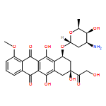
A novel hyaluronic acid modified transfersome was prepared to deliver drugs to lymphatics through the transdermal route. Doxorubicin loaded HA-GMS-T was able to efficiently penetrate into the deep skin tissue, leading to enhanced absorption by lymphatics. Most importantly, hyaluronic acid effectively improved the uptake of drug loaded nanocarriers by tumor cells.
Co-reporter:Yuanyuan Gao, Xiaojie Cheng, Zhiguo Wang, Juan Wang, Tingting Gao, Peng Li, Ming Kong, Xiguang Chen
Carbohydrate Polymers 2014 Volume 112() pp:376-386
Publication Date(Web):4 November 2014
DOI:10.1016/j.carbpol.2014.05.026
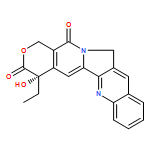
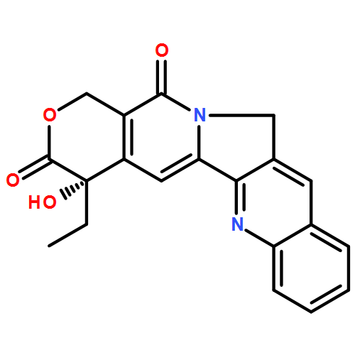
•HANs transferred MD-CPT into keloid lesion area and performed desirable skin permeable capacity.•MD-CPT/HANs was endocytosed by keloid fibroblast, played a dose-dependent inhibitory effect to KF.•They arrested cells at G1/S and prevented them entry into mitosis.•MD-CPT induced the significantly down-regulation of PAI-1 and up-regulation of Smad7.This study designs an alternative transdermal delivery system for 10,11-methylenedioxycamptothecin(MD-CPT) to inhibit keloid. Hyaluronic acid nanoemulsions (HANs) with nano size, negative charge and good stability were prepared as transdermal carriers. The MD-CPT loaded HANs performed desirable skin permeable capacity across human keloid skin and the drug was transferred directly to keloid lesion area. MD-CPT was delivered percutaneously higher than the control group. FITC-HANs could be successfully internalized by keloid fibroblast (KF) and deliver MD-CPT toward nucleus, inhibited the proliferation of KF, while there was no serious toxicity to normal skin fibroblasts. The growth-inhibitory effect was further clarified upon cell cycle regulation, which arrested cells at G1/S and prevented them entry into mitosis. KF gene expression demonstrated plasminogen activator inhibitor-1 (PAI-1) was significantly down-regulated and Smad7 up-regulated, which was beneficial to inhibit keloid. The study demonstrated that as transdermal delivery of MD-CPT by HANs has potential for inhibition of keloid fibroblast.
Co-reporter:Ming Kong, Hyunjin Park, Chao Feng, Lin Hou, Xiaojie Cheng, Xiguang Chen
Carbohydrate Polymers 2013 Volume 94(Issue 1) pp:634-641
Publication Date(Web):15 April 2013
DOI:10.1016/j.carbpol.2013.01.091
To develop a functional nanosized transdermal drug delivery system for tumor therapy, amphiphilic hyaluronic acid (HA) based niosome was constructed combining transdermal and tumor targeting ability in one entity. HA esterified with monostearin, the conjugate labeled as HA–GMS self-assembled onto niosome surface and formed HA–niosome. The multilayer vesicle had small size (around 40 nm), good stability and desirable drug encapsulating efficacy, and well compatible with blood. It exhibited better endocytosis to mouse breast tumor cell (4T1) than the control chitosan nanoparticle, which was verified qualitatively and quantitatively. Skin permeation of HA–niosome was proven to be efficient using in vitro stratum corneum model and in vivo fluorescence observation. Histological section study confirmed the security and efficiency of transdermal permeation. The results evidence HA–niosome to be exciting and promising for tumor therapy through trandermal administration.Highlights► Hyaluronic acid niosome as functional transdermal carrier for tumor therapy. ► HA–niosome has small size, elastic multilayer structure and hydrophilic interface. ► The incorporation of HA promotes the endocytosis of nanocarrier by tumor cell. ► HA–niosome is efficient and secure for transdermal permeation. ► HA–niosome is potential carrier for tumor therapy by percutaneous administration.
Co-reporter:Ming Kong, Lin Hou, Juan Wang, Chao Feng, Ya Liu, Xiaojie Cheng and Xiguang Chen
Chemical Communications 2015 - vol. 51(Issue 8) pp:NaN1456-1456
Publication Date(Web):2014/12/08
DOI:10.1039/C4CC08746A
A novel hyaluronic acid modified transfersome was prepared to deliver drugs to lymphatics through the transdermal route. Doxorubicin loaded HA-GMS-T was able to efficiently penetrate into the deep skin tissue, leading to enhanced absorption by lymphatics. Most importantly, hyaluronic acid effectively improved the uptake of drug loaded nanocarriers by tumor cells.

![(7S)-7-ethyl-7-hydroxy-10H-1,3-Dioxolo[4,5-g]pyrano[3',4':6,7]indolizino[1,2-b]quinoline-8,11(7H,13H)-dione](http://img.cochemist.com/ccimg/135500/135415-73-5.png)
![(7S)-7-ethyl-7-hydroxy-10H-1,3-Dioxolo[4,5-g]pyrano[3',4':6,7]indolizino[1,2-b]quinoline-8,11(7H,13H)-dione](http://img.cochemist.com/ccimg/135500/135415-73-5_b.png)